Next gen scanner could unlock new treatments for brain injuries
The scanner combines PET/MRI technology for better preclinical imaging.

Northeastern University psychology and pharmaceutical sciences professor Craig Ferris has spent decades doing groundbreaking research using brain imaging.
Now, a new device will help him unlock even further developments, including new treatments for brain injuries
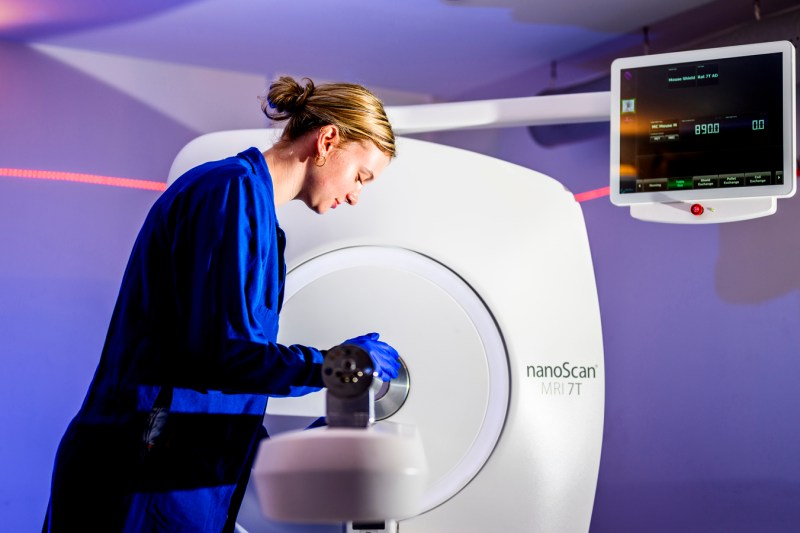
Ferris’ lab recently obtained a Mediso nanoScan 7T PET/MRI scanner, the first of its kind in the United States. The machine combines PET and MRI technology, allowing researchers to do both scans at once for better preclinical imaging.
“I don’t know of any combined magnetic resonance imaging and PET imaging being done in preclinical research in the Boston area,” says Ferris, whose research focuses on behavioral neuroscience and disease progression in psychiatric and neurological disorders. “It’s better technology than what we presently have. It’s the first of its kind, combining MRI technology with PET imaging.”


MRIs provide images of onsoft tissue structure, while PET scans use tracers that attach to a certain part of the body to show its function. What distinguishes this device is that it combines both functions to allow scientists to view different aspects of the brain at the same time.
“MRI is super good at soft tissue imaging while PET can tell you the functional part of (the brain),” said Praveen Kulkarni, a principal researcher in Northeastern’s psychology department. “Until now PET scans were always combined with CT scans. There is some equipment where they have a separate PET machine and separate MRI machine. But this is a combination of technologies into one single machine. That is what makes it unique.”
And Northeastern University was chosen to be the first place to showcase this technology in North America.
The new device comes from Mediso Medical Imaging Systems, an international company based in Hungary. Istvan Szanda, director of Mediso’s North American operations, said they were drawn to Northeastern due to Ferris’ research.


“Craig is a widely renowned professor in neuroscience,” Szanda says. “He hosted an educational session on MRI and one of our reps was drawn to the work and research of Craig and Northeastern. And we appreciate that Northeastern is strategically located within Boston and its biotech infrastructure. It’s critically important to showcase this system and further increase the prestige of Northeastern around biotechnology and pharmacology.”
In particular, Ferris’ work grabbed attention because he performs brain imaging on animals that are awake. Many other labs use anesthesia on the animals they’re studying in their preclinical research. Ferris prefers to study animals that are awake to mimic the state of the human brain.
“What’s going to happen here is truly unique,” added Gergo Bagamery, director of preclinical research and development at Mediso’s headquarters in Budapest. “We’ve worked in the past with academic sites on different projects, and what we understood is that the knowledge of how to use this imaging is really here.”
The lab will use the new scanner to look at both brain function and anatomy at the same time in order to better understand how the brain works under different conditions.
“Most of our research related to the brain and brain-related diseases,” Ferris said. “Our research goes from concussions to Alzheimer’s. Now we can combine brain function and anatomy with molecular biology. This machine allows another modality that gives you a whole sense of the mechanism behind how the brain is changing its activity.”
One example of how it can be used is to help develop new treatments for brain injuries. Ferris added that his lab is working with companies that develop psychedelics as a means of treatment for brain injuries, but without the hallucinogenic component. This new device will allow researchers to identify the receptors that receive that therapeutic part of the drug, allowing companies to refine the treatment.
The same can be done for puzzling out treatments for diseases like Parkinson’s disease or glioblastoma.
The new equipment is also less cumbersome than the 18-year-old PET scanner Ferris’ lab currently uses. Unlike its predecessor, the nanoScan 7t PET/MRI scanner can be unplugged and plugged back in easily, making it more “green” and easy to maintain. The current technology the lab has must be kept running at all times and is expensive to maintain.


